Mitalipov: Mitochondrial replacement therapy (2016)
OHSU offers select multimedia assets for use by news outlets. (Commercial use is prohibited without prior written consent.)
When using these materials, please:
-
Credit photos and video with “OHSU/Photographer name.” Photographer name, caption and usage restrictions are embedded in the image metadata. Do not alter the content/nature of the photo. Color correction and cropping are allowed.
-
Audio and video clips should be credited to OHSU.
For more information, please contact OHSU Strategic Communications at news@ohsu.edu or 503-494-8231.

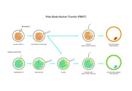
Mitochondrial replacement therapy
A pipette pulls out the nuclear genetic material from an unfertilized egg –a key step in mitochondrial replacement therapy. The nucleus, or spindle, is visible in the pipette. (OHSU)

ST in human oocytes
Shoukhrat Mitalipov's spindle-transfer technique. (OHSU)

Shoukhrat Mitalipov, Ph.D.
Shoukhrat Mitalipov, Ph.D., director of the OHSU Center for Embryonic Cell and Gene Therapy. (OHSU/Jeffrey Ball)

Hong Ma, M.D., Ph.D.
Hong Ma, M.D., Ph.D., staff scientist responsible for the management of the stem cell and molecular biology lab within OHSU’s Center for Embryonic Cell and Gene Therapy. (OHSU/Jeffrey Ball)

Mitochondria
Confocal microscopy image of Dr. Shoukhrat Mitalipov's work with human fibroblasts derived from —SCNT-ESCs. Nuclei are show in blue; mitochondria in red; and microtubules appear in green. (OHSU)

Mitochondria
A magnified section of a confocal microscopy image from Dr. Shoukhrat Mitalipov's work, showing human fibroblasts derived from —SCNT-ESCs. Nuclei are show in blue; mitochondria in red; and microtubules appear in green. (OHSU)

Mutant-iPSCs
Image from Dr. Shoukhrat Mitalipov's work, showing a human iPS cell colony. (OHSU)
- Latest Stories
- Popular


